
Discover clinical studies, expert guidance, and industry trends on advanced skin imaging and aesthetic diagnostic technology.
The 2026 market for Fluorescence light skin scanner technology is becoming more specialized. Clinics now compare ultraviolet fluorescence systems, blue-light scanners, multispectral platforms, and AI-assisted imaging devices. Each type reveals different visual signals, including surface oil, pigment patterns, hydration changes, and hidden pore congestion. However, these signals are not medical diagnoses.
Dr. R. Rox Anderson, a leading authority in laser medicine and dermatologic technology, has expressed a useful principle: “A device is only as useful as the question it answers.” This idea matters when selecting a Fluorescence light skin scanner. A bright image may look impressive, but image quality, calibration, operator training, and clinical interpretation matter more. Experienced practitioners examine the patient’s skin under consistent lighting, record baseline images, and compare changes over time.
The strongest scanners do not simply produce colorful maps. They support repeatable observation. Some systems use narrow-band blue illumination to highlight bacterial-related fluorescence. Others combine ultraviolet and visible wavelengths for broader surface analysis. Multispectral models can offer richer data, but they may require more training and higher maintenance costs. AI features can improve organization, yet they should not replace professional judgment.
The ranking is not perfectly settled.
A practical buyer should question exaggerated claims, request validation data, and inspect real workflow details. Can the scanner capture stable images on different skin tones? Does it protect patient privacy? Can clinicians explain the results clearly? These questions make the 2026 comparison more useful, realistic, and trustworthy.
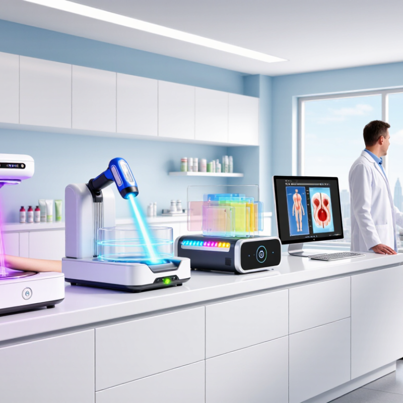
Fluorescence light skin scanners are imaging devices that reveal skin features invisible under ordinary lighting. They usually combine ultraviolet or blue excitation light with optical filters and a camera. Certain substances in skin emit visible fluorescence after excitation. Sebum, porphyrins, keratin, and some pigments can create different colors or intensities.
The scanner captures these signals and converts them into a surface map. Bright red-orange areas may suggest porphyrin activity, while pale or uneven fluorescence can indicate dryness, residue, or pigmentation changes. Results depend on distance, room light, skin cleansing, and camera calibration.
The American Academy of Dermatology states that professional skin evaluation remains essential because imaging cannot replace clinical diagnosis. That limitation matters. A striking image can still mislead.
Grand View Research valued the global skincare market at about USD 155.7 billion in 2023, with continued growth expected through 2030. This expansion supports wider interest in measurable skin analysis, but market growth does not prove scanner accuracy. In practice, operators should compare images under consistent conditions and record exposure settings.
Tips: Cleanse the face gently before scanning. Remove makeup, sunscreen, and excess oil. Keep the camera at a fixed distance. Avoid treating one fluorescent pattern as a confirmed condition. Repeat scans under matching settings. Some findings remain uncertain. Human review is still necessary.
In 2026, fluorescence light skin scanners are mainly classified by design, light control, and workflow. Handheld scanners suit quick checks in clinics, salons, and training rooms. They are portable and easy to position around the cheeks, forehead, or jawline.
Tabletop scanners provide steadier distance and lighting. This can improve image consistency during repeated assessments. Larger imaging systems capture the whole face under controlled conditions. They often support side-by-side comparisons and detailed skin mapping. They capture more.
Another important type uses controlled violet or blue excitation light to reveal visible fluorescence. Single-band scanners are simpler and usually easier to operate. Multispectral scanners use several light ranges, helping professionals examine different fluorescence responses. Some systems combine fluorescence images with surface photographs. This pairing can show texture, redness, or uneven tone beside the fluorescence pattern.
However, fluorescence results may change with cosmetics, moisture, room lighting, and camera distance. No category is perfect. Calibration still matters, even with advanced software.
Experienced operators should avoid treating a bright area as a confirmed medical condition.
Tips: Clean the skin gently before scanning, and remove reflective makeup. Keep the room dim and use the same distance each time. Record the device settings and scan angle. A scan is not a diagnosis. Suspicious or changing findings need review by a qualified healthcare professional. Also, compare results carefully; attractive images can create false confidence.
2026 Top Types of Fluorescence Light Skin Scanners
Fluorescence scanning uses controlled blue or ultraviolet light to reveal patterns hidden under normal lighting. Porphyrins, produced by certain skin bacteria, may appear as orange-red spots around pores. These signals can suggest acne-prone areas, but they cannot confirm an infection or medical diagnosis.
The images may also highlight excess sebum, follicular buildup, flaky surface cells, and uneven cleansing. Some scanners compare brighter and darker zones across the forehead, nose, cheeks, and chin. This helps professionals discuss oil balance and congestion with clearer visual evidence. Small details matter.
Pigment irregularities may look more noticeable under specialized illumination, especially when surface texture changes light reflection. Dry patches can appear uneven, although fluorescence alone cannot measure hydration precisely. A trained practitioner should combine the scan with skin history, touch, and visible examination. Not a diagnosis.
Results can change with recent cleansing, cosmetics, strong lighting, or camera distance. This is where interpretation becomes imperfect. A bright spot may reflect residue rather than a skin condition. Careful devices use consistent distance, exposure, and image settings. Qualified users should explain uncertainty instead of promising dramatic results. The most useful feature is not a colorful image, but a repeatable record that supports safer, individualized skincare decisions.
Clinical, Cosmetic, and Research Applications
Fluorescence light skin scanners use selected wavelengths to reveal surface and subsurface signals. Clinical systems may highlight porphyrins, oil-related fluorescence, or uneven pigmentation. These findings can support examination, but they do not replace medical diagnosis. In practice, interpretation is rarely neat. Skin tone, cosmetics, room lighting, and camera distance can change the image. Trained professionals should compare scans with medical history and direct observation.
Cosmetic scanners often provide visual maps for hydration concerns, texture, redness, and product consultation. Their value is strongest when measurements are repeated under consistent conditions. A single bright area is not proof of infection or damage. Research-grade systems usually offer controlled illumination, spectral data, calibration tools, and exportable measurements. Researchers can study barrier changes, treatment responses, or ingredient effects with stronger reproducibility. Still, even advanced equipment can produce misleading patterns when protocols are poorly designed.
Tips: Clean the skin gently before scanning. Record lighting, distance, and exposure settings. Remove reflective products when appropriate. Use the same device and protocol during follow-up visits. Protect privacy, obtain informed consent, and store images securely. Review unusual findings with qualified clinicians. Do not promise results from fluorescence alone. Some signals remain difficult to interpret, and that limitation deserves honest attention.
2026 Top Types of Fluorescence Light Skin Scanners
Safety, Accuracy, and Selection Considerations
Fluorescence skin scanners commonly use ultraviolet, violet, or blue excitation light. Each method reveals different surface signals, including porphyrins, oil, dryness, and uneven texture. A bright fluorescent area is not automatically a disease sign. Safety comes first. The device should control light intensity and exposure time. Users should follow tested operating procedures and protect the eyes when required. Skin should also be clean, dry, and free from reflective cosmetics before scanning.
Accuracy depends on more than camera resolution. Ambient light, skin moisture, recent skincare, and natural skin tone can change the image. A reliable scanner needs stable calibration and repeatable results. It should perform consistently across different users and skin conditions. Experienced operators compare fluorescence images with visible-light views and relevant clinical observations. They do not rely on one glowing patch. That matters.
When selecting a system, check its excitation range, image quality, calibration process, and software transparency. Ask whether the device supports storage controls, operator training, and documented testing. A simple interface helps, but it cannot replace professional judgment. I would also test several scans under the same conditions before purchase. The results may look impressive, yet small inconsistencies can remain. No scanner is perfect. Selection should reflect the intended setting, staff expertise, maintenance capacity, and acceptable risk level.
| Scanner Type | Typical Excitation Range | Primary Fluorescent Signal | Common Clinical or Cosmetic Use | Accuracy Characteristics | Main Limitations | Safety Considerations | Best Selection Scenario |
|---|---|---|---|---|---|---|---|
| Wood's Lamp / Long-Wave UVA Viewer | Usually centered near 365 nm; long-wave UVA approximately 315–400 nm | Fluorescence from selected skin substances, scales, pigments, and microbial metabolites | Visual assessment of pigment contrast, some superficial infections, and surface changes | Useful as a screening aid; interpretation is mainly qualitative and examiner-dependent | Ambient light, cosmetics, skin tone, hydration, and instrument distance can change the appearance; not a stand-alone diagnostic test | UVA eye and skin exposure should be minimized; use shielding, short examination times, and follow applicable UV safety procedures | Low-cost preliminary examination and routine dermatology or aesthetic consultation |
| Blue-Light Fluorescence Imaging | Commonly approximately 400–470 nm, depending on the optical filter set | Visible fluorescence from porphyrin-related compounds and other endogenous fluorophores | Acne-related follicular assessment, oiliness screening, and educational skin imaging | Can provide repeatable images when exposure, distance, and white balance are standardized; fluorescence intensity is not equivalent to bacterial count | Signal may be affected by sebum, cosmetics, cleansing, camera exposure, and room lighting; false positives and false negatives are possible | Generally lower photobiological concern than UV excitation, but direct high-intensity viewing should still be limited and eye-safety labeling should be checked | Client-friendly visualization and standardized before-and-after documentation |
| UV-Induced Multispectral Fluorescence Scanner | Multiple bands, commonly including approximately 365, 405, and 450 nm | Combined signals from endogenous fluorophores, surface materials, and selected metabolic by-products | Skin-surface mapping, lesion documentation, treatment monitoring, and research imaging | Higher information content than single-band viewing; quantitative reliability depends on calibration, reference targets, and validated algorithms | More expensive and complex; spectral overlap and biological variability can make component identification difficult | UV channels require exposure controls, shielding, and documented operating procedures; the device should have verified electrical and optical safety compliance | Clinics or research settings requiring image comparison across several excitation wavelengths |
| Porphyrin-Optimized Facial Fluorescence Camera | Usually violet or blue excitation near 405 nm, with a camera filter for red-orange emission | Red-orange fluorescence associated mainly with porphyrin compounds on or near pilosebaceous units | Acne-prone area visualization and progress tracking during skincare programs | Good for locating relative fluorescence patterns under controlled conditions; it does not directly measure acne severity or prove infection | Results vary with cleansing, sebum level, recent product use, camera geometry, and image-processing settings | Use eye protection or controlled viewing when required; avoid unnecessary repeated exposure and follow the device's irradiance limits | Aesthetic centers that need consistent visual documentation of acne-related fluorescence patterns |
| Time-Resolved Fluorescence Imaging System | Typically uses pulsed or modulated excitation in the UV, violet, or blue range | Fluorescence lifetime or decay behavior of endogenous skin fluorophores | Research, tissue characterization, and advanced assessment of biochemical or structural changes | Less dependent on absolute brightness than basic imaging; potentially more specific, but requires validated protocols and trained interpretation | High cost, lower workflow simplicity, motion sensitivity, and limited comparability between systems without harmonized calibration | Pulsed optical exposure must remain within documented limits; operators should receive training in laser or intense-light safety where applicable | Universities, hospitals, and laboratories conducting validated imaging studies |
| Fluorescence Dermatoscopy Attachment | Usually visible blue or violet excitation; some systems may add UVA illumination | Localized fluorescence observed together with magnified surface morphology | Close-up examination of selected lesions, texture, scales, and surface deposits | Magnification improves documentation of small areas; fluorescence remains an adjunct and does not replace dermoscopic or histopathological assessment | Small field of view, contact pressure effects, focus variation, and operator-dependent interpretation | Disinfect contact components appropriately; control optical exposure and avoid direct viewing of intense illumination | Targeted examination when a clinician needs both magnified morphology and fluorescence information |
The main types are handheld, tabletop, full-face, single-band, and multispectral scanners.
Handheld scanners suit quick checks in clinics, salons, and training rooms. They can reach the cheeks, forehead, and jawline easily.
It keeps the scanning distance and lighting steadier. This can improve consistency during repeated assessments.
Larger systems capture the whole face under controlled conditions. They support side-by-side comparisons and detailed skin mapping.
Controlled light can reveal fluorescence patterns hidden under normal lighting. Orange-red spots may appear around pores.
No. They may suggest acne-prone areas, but they cannot confirm infection or any medical diagnosis.
Scans may show excess sebum, follicular buildup, flaky cells, uneven cleansing, and pigment irregularities.
Clean the skin gently and remove reflective makeup. Keep the room dim. Use the same distance and scan angle each time.
Yes. Cosmetics, moisture, recent cleansing, room lighting, and camera distance can change the images.
Use them with skin history, touch, and visible examination. A bright area may be residue, not a skin condition. Interpretation is imperfect.
A Fluorescence light skin scanner is a noninvasive imaging device that uses controlled ultraviolet or violet light to stimulate natural fluorescence in the skin. Different substances, such as oil, pigmentation, keratin, and certain surface microorganisms, may emit distinct colors or patterns, helping reveal features that are not easily visible under ordinary lighting. In 2026, common types include handheld scanners, desktop diagnostic systems, camera-based imaging devices, and integrated analysis platforms with software-supported reporting.
These scanners can assist in observing dryness, excess oil, uneven pigmentation, clogged pores, surface impurities, and changes associated with skin conditions. They are used in clinical assessment, cosmetic consultations, skincare monitoring, education, and research. However, fluorescence results should be interpreted alongside visual examination and professional judgment rather than treated as a standalone diagnosis. When selecting a device, users should consider light safety, image consistency, calibration, resolution, data privacy, ease of operation, and the quality of its analysis features. Proper operating procedures and suitable protective measures are essential for reliable and responsible use.