
Discover clinical studies, expert guidance, and industry trends on advanced skin imaging and aesthetic diagnostic technology.
Choosing the best digital facial skin imaging system in China requires more than comparing screen size or promotional images. The right choice depends on the clinic’s workflow, the questions practitioners need to explore, and the consistency of each scan. A Digital facial skin imaging device may capture visible features such as pores, spots, redness, and texture under controlled lighting. Some systems also provide other imaging modes, but their outputs should be understood as visual information, not a diagnosis. Details matter.
Buyers should examine how the camera controls lighting, distance, and facial positioning. Repeatable conditions can make before-and-after comparisons more useful. Ask for a live demonstration using different skin tones and realistic clinic conditions, then review how clearly the software labels and stores results. Check whether staff can explain the images in plain language. A polished report is not automatically a reliable one. Results may vary with setup, operator technique, and software settings, so ask about calibration, training, maintenance, and technical support in China. Request clear documentation of the device’s intended use and performance claims, rather than relying on a single impressive sample image. No device tells the whole story. This guide compares practical selection criteria and common trade-offs, while recognizing that no single model will suit every clinic. Careful evaluation helps buyers choose a system that supports informed conversations without overstating what an image can show.
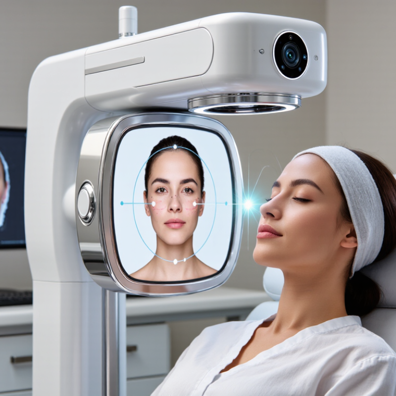
A digital facial skin imaging device captures a face under controlled lighting and analyzes visible skin features with imaging software. Depending on its setup, it may map pores, fine lines, pigmentation, redness, and surface texture. Some systems use cross-polarized or ultraviolet light to reveal details that ordinary room lighting can hide. The result is a visual record, not a medical diagnosis. It can help practitioners compare images over time, provided camera distance, lighting, and skin preparation stay consistent. Small changes in setup can distort the comparison. That matters.
For buyers in China, useful questions include whether the device produces repeatable images, explains its measurements, and offers staff training and dependable local support. Grand View Research estimated the global skin care devices market at USD 17.6 billion in 2023, with growth projected through 2030. This is broad market data, not proof that any imaging system is accurate. Ask for sample reports and repeat-scan results under the same conditions. Check how client images are stored and who can access them. Software can make skin look neatly quantified. Skin is still more complicated than a score.
A digital facial skin imaging system begins with a consistent setup. The user rests their chin on a support while the camera captures the face under controlled lighting. Multiple light modes can reveal different surface details. Cross-polarized light reduces glare, while other wavelengths may help show pigmentation patterns or redness. This matters. A small shift in head position or room lighting can change the image, even when the skin has not changed.
Software then maps visible features, such as pores, spots, fine lines, and uneven tone. It may compare left and right facial areas or track images taken during later visits. For useful tracking, photos should use similar lighting, distance, and facial expression. Results are estimates, not a medical diagnosis. The boundary between a visible feature and a software category is not always neat. Still imperfect. Dryness, recent sun exposure, or makeup residue can affect readings. An experienced operator should review the images, explain their limits, and avoid treating a score as the whole story. A photograph can guide discussion, but it cannot capture every cause of a skin concern.
Best Digital Facial Skin Imaging Device in China?
Which Criteria Matter When Comparing Devices in China?
Compare repeatability before counting features. Ask each supplier to scan the same person three times, then compare redness, pore, and pigmentation maps. The room, camera distance, facial position, and lighting should stay fixed. Otherwise, a lighting shift may look like a skin change. A crisp image can still mislead.
Check whether the system performs consistently across different skin tones and ages. NIST’s 2019 Face Recognition Vendor Test assessed 189 algorithms and found demographic differences in false-positive rates, sometimes ranging from 10 to 100 times. This tested face recognition, not skin analysis, so it cannot prove a skin scanner’s accuracy. Still, it highlights why vendors should show evaluation results across varied faces, not only ideal test images.
For clinics in China, examine practical workflow: local-language reports, clear explanations for clients, and export options that fit existing consultation routines. Ask how image data is stored, who can access it, and how long retrieval takes. Request sample reports and repeat-scan results before purchase. Small details matter. A confusing color scale can undermine a technically strong device, and I would not judge performance from polished demonstrations alone.
| Comparison criterion | Standard 2D RGB imaging | Multispectral or polarized imaging | 3D facial imaging | What to check before purchase |
|---|---|---|---|---|
| Image information | Visible-light color photographs show surface appearance, such as visible redness, spots, and texture. | May add selected illumination or polarization modes to reveal features that are less apparent in a standard color image. Available modes vary by device. | Captures facial shape and depth information in addition to the visible image. | Confirm which image modes are actually included and what each mode is designed to measure. |
| Common assessment uses | Before-and-after photographs and visual review of visible skin features. | Comparative review of selected skin features under different lighting or polarization conditions. | Facial surface topography, contour, and dimensional comparison. | Match the device’s documented functions to the clinic’s intended assessment or follow-up workflow. |
| Capture consistency | Depends on consistent lighting, camera position, distance, and facial expression. | Also depends on consistent positioning and the correct use of each illumination mode. | Requires repeatable positioning and capture conditions for meaningful shape comparisons. | Look for positioning guides, fixed capture settings, and clear repeat-capture instructions. |
| Lighting and environment | Ambient light and reflections can affect the appearance of color images. | Controlled illumination is important; reflections and inconsistent capture conditions may affect comparisons. | Lighting can affect the texture and color view, while depth capture depends on the system’s sensing method. | Ask whether the system controls or checks lighting and whether the room needs specific conditions. |
| Repeatability | Best assessed using the same camera setup, settings, and positioning across visits. | Depends on repeatable acquisition for every included mode, not just the standard image. | Depends on consistent facial alignment and the device’s depth-measurement process. | Request repeatability information and a demonstration using repeated captures under the same conditions. |
| Analysis and scores | Some systems provide image comparison or automated analysis; features differ by software. | Analysis may be available for selected modes, but interpretation and scoring differ by system. | May provide measurements or visualizations of facial depth and surface shape; tools vary. | Ask how each score is calculated, what it represents, and whether supporting validation information is available. |
| Clinical interpretation | Images support visual documentation but do not, by themselves, establish a medical diagnosis. | Additional image modes provide information for review but should not be treated as a diagnosis on their own. | Shape measurements can support comparison but do not independently establish a diagnosis. | Check intended use, user guidance, and the limits of automated interpretation. |
| China-specific suitability | Image capture capability is separate from local software, service, and compliance arrangements. | Additional imaging modes do not by themselves confirm local regulatory status or service availability. | 3D capability does not by itself confirm local regulatory status or compatibility with clinic systems. | Verify applicable China regulatory requirements, Chinese-language support, data handling, training, warranty, and local maintenance with the supplier. |
| Data management | Check image storage, export formats, access controls, and backup options. | Check whether all image modes can be stored, retrieved, and exported together. | Check storage and export options for both images and 3D data. | Review where data is stored, who can access it, retention settings, and applicable privacy obligations. |
| Practical selection | May suit workflows centered on standardized visible-light photographs. | May suit workflows needing additional controlled imaging modes, depending on the specific configuration. | May suit workflows that require facial shape or depth comparison. | Compare actual specifications, demonstration results, operating costs, service terms, and workflow fit—not category labels alone. |
Comparison note: These are general technology categories, not guaranteed specifications for every device. Features, measurement methods, intended uses, and performance vary by model and configuration. Request the device’s current technical documentation and verify local regulatory and data-protection requirements before purchase.
In China, skin imaging devices commonly fall into four groups: polarized-light systems, multispectral or UV analyzers, 3D facial scanners, and handheld dermatoscopes. Polarized light reduces surface glare, making pores, redness, and uneven texture easier to inspect. UV and multispectral systems estimate pigment and oil patterns beneath the visible surface. 3D systems map facial contours and track volume changes. Handheld scopes offer close views of individual spots.
Small differences matter.
The International Society of Aesthetic Plastic Surgery’s 2023 Global Survey reported about 19.1 million non-surgical aesthetic procedures worldwide. That figure is not a scanner-sales count, but it signals a large setting for visual assessment. In Chinese clinics, device choice often depends on the task: broad complexion screening, treatment tracking, or examination of one lesion.
Ask how images are captured, whether lighting stays consistent, and what the software actually measures. A colorful score is not a diagnosis. Performance can vary with skin tone, makeup, and room lighting; this deserves more attention than polished sample images usually receive.
For suspicious lesions, imaging should support—not replace—a qualified clinical examination. The practical fit can be less impressive than the brochure.
Choosing a facial imaging system starts with the clinic’s workflow, not its most impressive sample image. A busy practice may need quick capture, clear side-by-side comparisons, and simple patient reports. A specialist clinic may prioritize consistent lighting, cross-polarized views, and detailed pigmentation or texture maps. Check how long setup takes, how staff learn the software, and whether images remain comparable across visits. Small details matter.
Demand is growing, but procedure counts do not prove that every clinic needs the same device. The American Society of Plastic Surgeons’ 2023 Procedural Statistics Report recorded about 25.4 million minimally invasive procedures in the United States. ISAPS’ 2023 Global Survey reported about 15.8 million nonsurgical procedures worldwide; the figures use different scopes. Use them as context, not a purchasing forecast. Ask vendors for repeatability data and test images under your own room lighting and patient mix. Even a good setup has limits. A device supports assessment and communication; it should not replace clinical judgment. Also compare service response times, staff training, storage options, and total operating costs. Reality is messier: inconsistent positioning or a changed light source can make follow-up images misleading.
The user rests their chin on a support while a camera photographs the face under controlled lighting. Different light modes reveal different details. Cross-polarized light reduces surface glare.
Software may map pores, spots, fine lines, redness, and uneven tone. It can compare facial areas or track images across visits. Results are estimates, not a diagnosis.
Common types include polarized-light systems, multispectral or UV analyzers, 3D facial scanners, and handheld scopes. Each serves a different purpose. Some show broad patterns; others magnify one spot.
Keep lighting, camera distance, head position, and facial expression consistent. A changed lamp or tilted chin can make images look different. The skin may not have changed.
No. A photograph can support a discussion, but it cannot show every cause. Treating a score as the whole story is tempting, but unwise.
No. Imaging supports assessment but does not replace qualified clinical judgment. Suspicious spots need proper examination. A polished image is not enough.
Makeup residue, dry skin, recent sun exposure, skin tone, and room lighting can affect readings. Small details matter. They are easy to overlook.
Match the device to the clinic’s workflow and assessment needs. Test images under your own lighting and with your patient mix. Ask about repeatability, staff training, storage, service, and operating costs. The brochure may look better than daily use.
A Digital facial skin imaging device uses specialized cameras and lighting to create detailed images of the face, helping clinics observe visible skin features such as pores, pigmentation, redness, texture, and fine lines. By capturing images under consistent conditions and analyzing them with imaging software, these systems can support consultations, document changes over time, and help practitioners explain skin-care options. Results should be interpreted alongside a professional assessment rather than treated as a diagnosis on their own.
When comparing devices in China, clinics can consider image clarity, analysis functions, ease of use, report quality, data handling, training, maintenance, and overall cost. Available options range from basic imaging tools for routine visual records to more advanced systems with multiple lighting modes and expanded analysis features. The most suitable choice depends on a clinic’s services, patient volume, budget, and workflow. Evaluating these practical needs can help clinics select a system that fits their environment and supports consistent, clear communication with patients.