

Blog & Insights
Discover clinical studies, expert guidance, and industry trends on advanced skin imaging and aesthetic diagnostic technology.
Discover clinical studies, expert guidance, and industry trends on advanced skin imaging and aesthetic diagnostic technology.
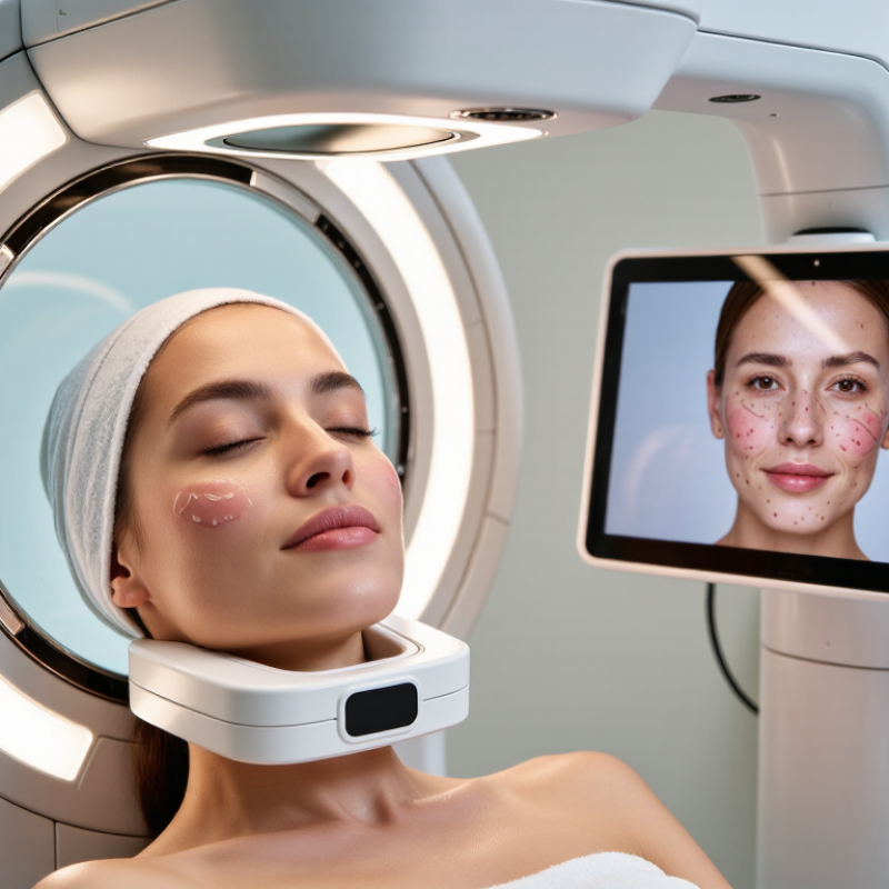
A Professional beauty skin imaging system can reveal details that a mirror and ordinary room lighting often miss: uneven tone, visible pores, fine lines, and changes across the cheeks or forehead. Yet a vivid image is not a diagnosis. The American Academy of Dermatology recommends personalized skin care, and imaging is most useful when trained professionals interpret results alongside client history and direct observation. In practice, consistent lighting, repeatable positioning, and clear explanations matter as much as the screen’s impressive colors.
The commercial interest is growing. Grand View Research estimated the global skin care market at US$115.65 billion in 2024, reflecting a large and competitive beauty sector—not proof that any particular imaging device improves outcomes. Buyers should compare image consistency, measurement transparency, staff training, privacy safeguards, and ongoing service, rather than choosing by feature count alone. Dermatologist Dr. Zoe Diana Draelos is a recognized authority in cosmetic dermatology; however, without a verifiable source for a direct quotation about professional skin imaging, assigning her a quote would be misleading. That gap matters. This guide examines what the available technology can show, where its limits lie, and how to assess systems with informed skepticism.
What Is the Best Professional Beauty Skin Imaging System?
How Professional Skin Imaging Systems Capture Standardized Facial Images
A professional skin imaging system should make repeat photographs comparable, not merely impressive. The American Society of Plastic Surgeons’ 2023 procedural report recorded about 25.4 million minimally invasive procedures in the United States, highlighting the value of consistent visual records in aesthetic care. That figure measures procedures, not imaging-system performance.
Standardization begins before the shutter clicks. The camera stays at a fixed height and distance, with consistent exposure, a neutral background, and controlled, diffuse lighting. The client faces forward, then turns to matching angles; hair is moved away from the face, and expression remains relaxed. Some systems also use cross-polarized lighting to reduce surface glare and reveal details beneath it. These images can support consultation and progress tracking, but they do not replace clinical assessment. One limitation deserves attention: even controlled lighting may render different skin tones unevenly.
Tips: Mark the chair and camera positions on the floor. Keep room lighting consistent, and record the settings. Review a test image for glare, shadows, and color shifts before capturing the full set. Standardization takes practice. A rushed setup can quietly undermine the comparison.
| System type | How images are captured | Typical image outputs | Useful for assessing | Key considerations |
|---|---|---|---|---|
| Standardized 2D facial photography | A camera, fixed subject position, controlled lighting, and repeatable front and angled views are used to capture visible-light photographs. | Color facial images from consistent views; some systems also provide side-by-side comparison tools. | Visible changes in complexion, blemishes, redness, and overall appearance when capture conditions are kept consistent. | Lighting, camera settings, facial expression, head position, and distance should be controlled for useful comparisons. |
| Cross-polarized imaging | Polarizing filters on the light source and camera reduce surface reflections in the captured image. | Images with reduced glare that can make some color variations easier to inspect. | Visible redness and pigmentation patterns that may be obscured by shine in ordinary lighting. | It changes how reflected light is recorded; results are not a direct diagnosis or a substitute for clinical assessment. |
| Ultraviolet photography | A UV light source and a camera configured for the relevant wavelengths capture reflected or emitted light from the skin. | UV images that may reveal patterns not readily visible in standard visible-light photographs. | Certain surface features and pigmentation patterns, depending on the imaging method and equipment. | UV imaging methods differ, and image appearance can be affected by wavelength, filters, and exposure. Use appropriate eye and skin safety measures. |
| Multispectral imaging | Images are captured using multiple selected wavelength bands, which may include visible light and other bands depending on the device. | Separate spectral images or derived visual maps for comparing selected image features. | Patterns such as visible pigmentation or redness, subject to the system’s validated methods and intended use. | The number and range of bands vary. Derived maps are image analyses, not automatically medical diagnoses. |
| 3D facial imaging | Multiple views or projected patterns are used to reconstruct facial surface shape and depth. | A three-dimensional surface model and measurements of facial geometry, depending on the system. | Facial contours and surface-shape comparisons over time. | 3D shape data is different from color or spectral skin analysis; many 3D systems do not provide UV or multispectral images. |
| Integrated imaging platform | Combines two or more capture modes, such as standardized color photography with polarized, UV, multispectral, or 3D imaging. | A set of aligned image types and comparison features; exact outputs vary by configuration. | Broader visual documentation when multiple image modes are relevant to the consultation. | Confirm which modes are included, how images are standardized, and whether image analysis is validated for the intended use. |
A suitable professional system should provide repeatable lighting and positioning, consistent image views, secure image storage, and clear comparison tools. The best choice depends on the intended use; imaging results support visual assessment and do not by themselves diagnose skin conditions.
It can show spots, wrinkles, texture, pores, UV patterns, brown spots, redness, and porphyrins. It does not diagnose medical conditions.
Polarized lighting reduces glare and helps show uneven tone, redness, pores, and fine texture. Small details become easier to inspect.
UV images may reveal hidden pigmentation, sun exposure patterns, or fluorescence linked with oil and porphyrins. These images are screening aids, not proof.
Cleanse gently and remove makeup, sunscreen, and heavy skincare products. Keep the room temperature steady.
Changes in distance or angle can make pores and other features look different. Matching each session makes comparisons more useful.
Redness and skin appearance can change after exercise, stress, spicy food, or irritation. Trends over several weeks offer better context.
Yes. Images can look dramatic, and rushed interpretation may cause unnecessary concern. A trained professional should explain both their value and limits.
Get clear consent before storing images and protect access to them. Record the date, settings, and relevant treatment history.
Imaging cannot confirm a diagnosis. A professional can explain what the images show and suggest clinical advice when appropriate.
A Professional beauty skin imaging system is designed to capture consistent, standardized facial images that help professionals evaluate skin condition more objectively. By controlling factors such as lighting, positioning, facial expression, and camera distance, these systems make it easier to compare results over time. A comprehensive assessment may examine eight common features: spots, wrinkles, texture, pores, UV-related concerns, brown spots, redness, and porphyrins. Different lighting methods provide different insights, with polarized light emphasizing surface details, redness, and texture, while UV illumination can reveal underlying pigmentation patterns and other features not easily seen under normal light.
Image interpretation should also consider Fitzpatrick’s six skin phototypes, since pigmentation and light response can vary significantly between individuals. When comparing systems, professionals should focus on image repeatability, reliable calibration, measurement consistency, and ease of integration into the clinical workflow. The best choice is not simply the system with the most visual effects, but one that produces dependable data, supports clear consultations, and helps track skin changes accurately and responsibly.