
Discover clinical studies, expert guidance, and industry trends on advanced skin imaging and aesthetic diagnostic technology.
Skin assessment has moved beyond the magnifying lamp and paper chart. Today, a Non-invasive skin analysis scanner can capture close-up images, map visible skin features, and turn them into reports for consultations or product planning. For clinics, salons, and skincare brands, the right system may save time and make changes easier to explain. Small details matter: lighting consistency, camera resolution, software workflow, and how clearly the results are presented.
This guide introduces ten manufacturers and compares the features buyers commonly examine, including imaging methods, report options, usability, training, and after-sales support. A polished screen is not enough. Ask how the device handles different skin tones, whether measurements can be repeated consistently, and what evidence supports its claims. Manufacturer specifications are useful, but they do not replace independent evaluation or qualified professional judgment. The category is not perfectly uniform, either; some systems focus on facial imaging, while others offer broader consultation tools. That distinction can be easy to miss.
There is no single best scanner for every setting. A busy skincare studio may value quick operation, while a research-oriented team may need deeper image controls and data handling options. Budget, service access, and staff training also shape long-term value. Even a strong system has limits. Skin changes with lighting, hydration, and daily routine, so scan results need context. Use this comparison as a practical starting point, then verify each manufacturer’s current specifications before making a purchase.
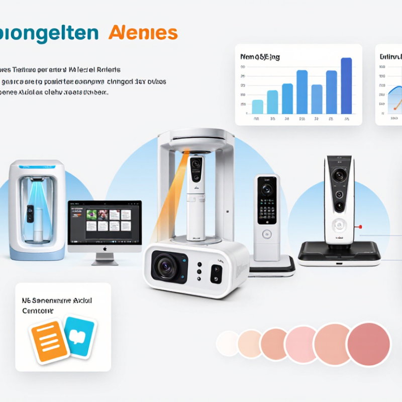
Non-invasive skin scanners use light, cameras, and contact sensors to record surface features without taking a sample. Standard photographs show visible tone and texture. Cross-polarized light can reduce surface glare, making redness and uneven pigmentation easier to inspect. Ultraviolet imaging may reveal spots not obvious under ordinary lighting.
Light matters.
Some systems combine several images with 3D measurements. Their software estimates pore visibility, wrinkle depth, spots, and facial symmetry. A contact-based sensor may assess electrical properties associated with skin moisture or oiliness. These are instrument readings, not medical diagnoses. Results depend on the device, its settings, and how the person is positioned.
For a useful comparison, keep lighting, camera distance, facial expression, and skin preparation consistent. A clinician or trained operator should explain what each score represents and whether it is an estimate or a direct measurement. Small changes may reflect a different angle, recent washing, or room humidity rather than a real skin change. Numbers can look more certain than they are. A limitation worth admitting: scanner scores do not capture every concern, and different systems may produce results that cannot be compared directly.
A useful skin analysis scanner combines four views, not four marketing filters. RGB imaging records visible tone, pores, and spots under controlled white light. UV imaging can reveal fluorescence patterns and features less apparent in ordinary photographs. Cross-polarized light suppresses surface glare, making redness and pigment contrast easier to inspect. 3D profilometry measures surface relief, including wrinkle depth and roughness. Light matters. Each modality answers a different question, and none alone describes overall skin health.
Commercial interest is growing: Grand View Research estimated the global skin care products market at USD 115.65 billion in 2024. The Global Wellness Institute put personal care and beauty at USD 1.09 trillion in 2022. These figures indicate a large consumer sector, not proof that a scanner’s measurements are clinically valid. For manufacturers, repeatability matters as much as image detail: fixed camera distance, consistent room lighting, clean lenses, and documented skin preparation help reduce noisy comparisons. Even a small change in exposure can make redness look stronger. Not a diagnosis. UV and cross-polarized images should be interpreted cautiously, with clear limits and trained review; attractive color maps can still oversimplify complex skin.
For non-invasive skin analysis scanners, repeatability matters more than a polished image. ISO 5725-1 defines repeatability under closely matched conditions, including the same method, operator, and equipment. In practice, a manufacturer should report repeated readings, sample size, and variation, not just a single best result. Small numbers matter. A 2% repeatability claim is hard to assess without knowing the test site, interval, and environmental controls.
Resolution should be stated as measurable detail, such as micrometres per pixel, and verified with a calibration target. Screen pixels alone do not prove skin-level resolving power. EEMCO guidance for transepidermal water loss (TEWL) recommends controlled testing conditions, commonly 20–22°C and 40–60% relative humidity, with acclimatization before measurement (Rogiers, Skin Pharmacology and Applied Skin Physiology, 2001;14:117–128). These details help explain why a reading can shift after a warm walk or a dry room.
TEWL should be reported in g/m²·h, alongside temperature, humidity, probe placement, and repeat-reading variation. Compare devices only when these conditions are similar. I would still question any “high accuracy” claim that omits its protocol; even careful measurements can drift across cheeks, foreheads, and testing days. A useful datasheet makes that uncertainty visible.
Performance metrics: repeatability, resolution, and TEWL (g/m²·h)
These metrics do not have a single universal cross-manufacturer benchmark that supports a fair numerical ranking. Repeatability depends on the test protocol, resolution is device-specific, and TEWL readings depend on measurement conditions and body site. Compare published specifications only when the methods and units are aligned; this chart does not represent manufacturer performance.
Comparing non-invasive skin analysis scanner manufacturers requires more than counting imaging modes. Multispectral cameras, cross-polarized light, and 3D capture reveal different features; they are not interchangeable. Ask for repeatability data across operators, lighting conditions, and skin tones, plus the reference method used for validation. A polished dashboard proves little. A 2017 Nature study trained a deep-learning model on 129,450 clinical images and compared its performance with 21 board-certified dermatologists. That research examined lesion classification, not cosmetic skin scoring, so it does not validate beauty scanners for diagnosis.
The distinction matters.
Compare validation by intended use: hydration tracking, pore visualization, pigmentation mapping, or clinical documentation. Request test-retest variation, sample size, and independent studies—not only manufacturer demonstrations. Check whether results stay stable after cleansing or changes in room temperature. Small details count. Visible-light imaging may suit consultation records, while multispectral systems can help distinguish redness from pigmentation; performance still depends on calibration and software. For everyday use, assess image export, staff training, maintenance, and whether scans can be repeated at a consistent distance. Reports with clear protocols are more useful than dramatic accuracy claims. Some specifications remain difficult to compare, and that limitation deserves attention.
When comparing the top 10 non-invasive skin analysis scanner manufacturers, look beyond image quality and feature lists. Ask whether the manufacturer operates an ISO 13485 quality management system, and verify that its scope covers the relevant design and production activities. This system supports controlled processes, traceable records, and corrective actions. It does not, by itself, prove that every scanner performs accurately. Request evidence for calibration, software validation, and image consistency under typical clinic lighting.
IEC 60601-1 addresses basic safety and essential performance for applicable medical electrical equipment. Check which edition and test configuration apply to the scanner. Review documentation on electrical insulation, leakage current, and safe operation, especially if the device connects to other equipment. Details matter. A certificate alone may not show how the finished scanner was assessed. Ask for test reports or a clear explanation of the assessment scope. Requirements can vary by intended use and market, so avoid assuming one document answers every question.
Tips: During a demonstration, capture the same skin area twice under fixed lighting and compare the results. Ask how the device flags poor-quality images and how updates are controlled. Check that service staff can trace calibration and repair records. A tidy dashboard is useful, but it cannot replace clear evidence. Even experienced buyers may find documentation uneven; note what remains unclear before making a shortlist.
The profiles below compare common scanner configurations, not named or verified manufacturers. They are not a market ranking. Performance and regulatory status vary by exact device and intended use; confirm claims against current model-specific documentation.
| Profile | Typical imaging configuration | Common non-invasive outputs | Useful benchmark questions | ISO 13485 quality-system check | IEC 60601-1 electrical-safety check | Interpretation boundary |
|---|---|---|---|---|---|---|
| Benchmark profile 01 | Standardized visible-light facial photography | Visible spots, redness, pores, wrinkles, and tone variation | Are lighting, camera distance, head position, and image capture repeatable? | Request a current certificate and confirm its scope covers the legal manufacturer and relevant production site. | Request the applicable test report or declaration for the exact model and configuration. | Image-based appearance assessment; outputs are not, by themselves, a medical diagnosis. |
| Benchmark profile 02 | Cross-polarized visible-light imaging | Surface redness and pigmentation patterns with reduced surface glare | Does the system disclose illumination conditions and keep capture settings consistent between visits? | Verify certificate validity, scope, and issuing body; do not infer certification from a product brochure. | Check model-specific electrical-safety evidence and any applicable national deviations. | Visual contrast can support comparison, but does not establish the cause of a skin finding. |
| Benchmark profile 03 | Ultraviolet illumination with compatible imaging | Fluorescence patterns and selected surface features visible under the stated illumination | Are wavelength, exposure controls, eye-safety instructions, and operating distance documented? | Confirm that the quality-system certificate applies to the organization responsible for the device. | Review electrical-safety evidence and the device instructions for illumination-related precautions. | Fluorescence appearance is not a definitive test for a particular condition or microorganism. |
| Benchmark profile 04 | Multispectral visible and near-infrared imaging | Comparative maps of selected pigmentation and redness-related image features | Are spectral bands, calibration methods, repeatability data, and limitations available? | Ask for the certificate number, scope, validity dates, and confirmation from the issuing body. | Confirm testing applies to the complete marketed system, including its power supply and accessories. | Mapped values depend on device algorithms and acquisition conditions; they are not interchangeable across systems. |
| Benchmark profile 05 | High-resolution macro or dermoscopic-style surface imaging | Magnified views of skin-surface structures and localized visible features | What is the stated optical resolution, field of view, and method for keeping scale consistent? | Verify the certified quality-system scope rather than relying on a general ISO claim. | Request applicable IEC 60601-1 documentation when the device is medical electrical equipment. | Magnified images require qualified interpretation where clinical assessment is intended. |
| Benchmark profile 06 | 3D optical facial imaging | Surface topography, contour visualization, and selected wrinkle or texture measurements | Are reconstruction accuracy, positioning repeatability, and measurement units stated? | Check certificate scope and whether the manufacturing and design activities are covered. | Review model-specific electrical-safety evidence, including the supplied power components. | 3D surface measurements describe captured geometry; they do not measure every underlying skin property. |
| Benchmark profile 07 | Polarized imaging combined with automated image analysis | Software-generated counts or scores for selected visible features | Are the scoring method, intended population, validation data, and version history disclosed? | Confirm that quality-system coverage includes relevant design and software lifecycle processes where applicable. | Check safety evidence for the physical device and identify which software functions are included. | Automated scores are method-dependent and should not be treated as clinical conclusions unless specifically authorized. |
| Benchmark profile 08 | Multi-angle facial image capture | Comparisons of visible features across frontal and angled views | Does the workflow control pose, expression, exposure, and image alignment between sessions? | Request current, independently verifiable documentation covering the relevant manufacturing entity. | Verify the applicable standard edition and evidence for the exact product configuration. | Changes in pose, lighting, or expression can affect image comparisons and derived scores. |
| Benchmark profile 09 | Portable camera-based skin imaging | Captured photographs and software-assisted tracking of visible changes | Are camera calibration, ambient-light limits, data handling, and capture guidance documented? | Confirm whether the portable device and its responsible manufacturer fall within the certificate scope. | Check the relevant electrical-safety documentation for the device, charger, and accessories. | Results may vary with the camera, lighting, and user technique; confirm intended use before clinical reliance. |
| Benchmark profile 10 | Combined imaging station with standardized acquisition software | Side-by-side image review, longitudinal comparisons, and selected feature summaries | Can the system export original images, identify software versions, and preserve an audit trail? | Verify certificate validity, scope, sites, and the legal entity responsible for the marketed system. | Request the exact-model test report or declaration and check whether accessories are covered. | Trend reports support documentation; they do not guarantee measurement equivalence or diagnostic accuracy. |
It records visible features such as tone, pores, spots, and texture. Some systems also estimate moisture or oiliness using contact sensors. These readings are not diagnoses.
RGB shows visible skin tone and texture under white light. UV may reveal patterns less obvious in ordinary photos. Cross-polarized light reduces glare, making redness and pigmentation easier to inspect. Light matters.
It measures surface relief, including wrinkle depth and roughness. A raised or shadowed area may look different under another camera angle, so the number needs context.
Keep the lighting, camera distance, facial expression, and skin preparation consistent. Use the same room and capture the same area twice. Small details matter.
Recent washing, room humidity, camera angle, or exposure settings can affect the readings. A change on screen may not mean the skin itself changed.
Not always. Systems may use different sensors, settings, and scoring methods. Their results can look precise, but they may not measure the same thing.
Ask whether it is an estimate or a direct measurement, and what the score represents. A trained operator should explain the limits. Not a diagnosis.
Ask whether an ISO 13485 quality system covers relevant design and production activities. For applicable electrical equipment, review IEC 60601-1 assessment details. Request calibration, software validation, and image-consistency evidence. A certificate alone may leave questions.
A Non-invasive skin analysis scanner uses optical imaging and related sensing methods to assess visible and measurable skin characteristics without damaging the skin. RGB imaging records surface tone and texture, UV imaging can reveal selected subsurface patterns, cross-polarized imaging helps reduce glare and highlight features such as redness, and 3D profilometry maps surface contours. Together, these modalities can support consistent documentation and help practitioners compare results over time.
When comparing the top 10 manufacturers, the article considers technology, validation, intended use, and practical performance rather than brand claims alone. Key measures include repeatability, image resolution, and, where separately measured, transepidermal water loss (TEWL), expressed in g/m²·h. It also explains why standardized testing matters: ISO 13485 quality systems relate to medical-device quality management, while IEC 60601-1 addresses electrical safety and essential performance. These benchmarks help readers assess whether a device is suitable for its intended setting and offers dependable, interpretable results.