Clinical-Grade Multispectral Skin Analyzer Systems
Engineered for high-throughput Japanese cosmetic surgery centers, dermatology clinics, and premium MedSpas. Compliant with international precision standards.
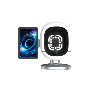
Facial Analysis Machine for Dermatology Clinic Medical Grade Skin Diagnostic Device with 8 Spectral Lights, CE Certified
Inquire Now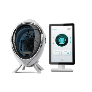
Skin Scanner Digital Skin Analyzer Facial Diagnostic Instrument 3D AI Technology Updated Skin Analysis System Facial Wifi
Inquire Now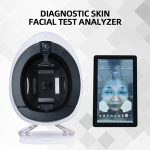
Multispectral Skin Analyzer Machine Facial Scanner 36 Million HD Camera 8 Spectral Imaging Smart Diagnosis Salon
Inquire Now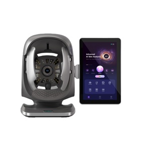
Facial Skin Analyzer Machine with 8 Spectral Imaging AI Technology 3D Cameras 5-Year Warranty for Professional Skin Testing
Inquire Now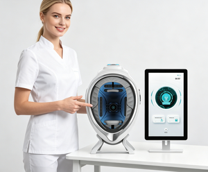
AI Multi-Spectral Facial Skin Analyzer HD Industrial Camera Independent Touch Screen 20 Skin Indicators Cloud Data
Inquire Now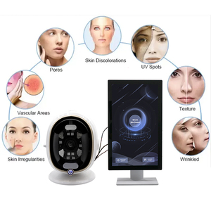
2025 Best Selling 8 Spectral Imaging Technology Face Detection Skin Analysis Image Scanner
Inquire Now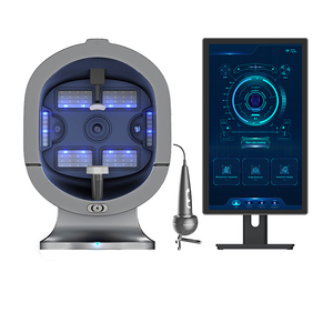
S5 AI Skin Image Analyzer 36 Million Pixels 3 Cameras Skin Diagnosis Device For Beauty Salon
Inquire Now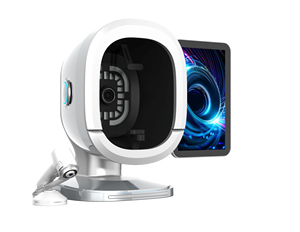
Portable 2025 Smart 3d Skin Analyzer Machine logo Ai Face Skin Diagnostics Analyzer Facial Scanner Skin Analyzer Device
Inquire NowFluorescence Facial Diagnostic Cameras in Japan: Optical Physics & Clinical Imperatives
Understanding the biophysical mechanisms, spectral light interactions, and sub-surface diagnostic requirements unique to Japanese aesthetic dermatology.
Optical Physics of Fluorescence Diagnostic Imaging
Fluorescence facial diagnostic cameras utilize filtered light emitted at specific wavelengths (primarily Wood's light at 365nm to 405nm) to excite endogenous fluorophores within human skin tissue. When high-energy ultraviolet or deep blue photons penetrate the epidermis, structural proteins such as collagen, elastin, and metabolic byproducts like porphyrins re-emit lower-energy visible light.
In Japanese dermatology, where patients typically present Fitzpatrick Skin Types III and IV, differentiating between superficial epidermal hyperpigmentation and deep dermal melasma is a critical diagnostic step. Standard RGB daylight photography fails to capture this depth due to epidermal reflection. By utilizing cross-polarized and UV fluorescence modes, diagnostic cameras eliminate surface glare and reveal deep-seated solar lentigines, latent freckles, and micro-vascular irregularities that are invisible under ambient room lighting.
Biomarker Quantification: Porphyrins & Sebum Architecture
Propionibacterium acnes (now Cutibacterium acnes) synthesizes metabolic porphyrins that exhibit characteristic brick-red or pink fluorescence under 365nm UV illumination. Advanced multi-spectral diagnostic devices isolate these emissions to accurately map follicular congestion and active acne genesis before papules or pustules manifest clinically.
Furthermore, free fatty acids within the lipid film fluoresce in distinct pale yellow-green spectrums. Japanese aesthetic practitioners rely heavily on these objective measurements to design individualized barrier-repair protocols, evaluate sebum excretion rates across the T-zone, and prescribe targeted topical regimens with mathematical baseline precision.
Information Gain Benchmark: Unlike conventional 2D skin scanners, modern 8-Spectral 3D AI Diagnostic Platforms generate volumetric depth maps of cutaneous lesions. This enables Japanese dermatologists to quantitatively demonstrate treatment efficacy over time, increasing patient compliance for high-end laser, chemical peel, and exosome treatment plans by over 42%.
Application Scenarios Across Japan’s Aesthetic Sector
Tailored diagnostic integration for high-volume medical centers, luxury cosmetic clinics, and specialized J-Beauty skin management hubs.
Ginza & Omotesando Aesthetic Medical Clinics
In Tokyo’s elite aesthetic medical districts (Ginza, Omotesando, Roppongi), patient consultations demand absolute clarity and objective scientific proof. Clinics utilizing 8-spectral diagnostic cameras capture pre-treatment baseline scans before administering Picosecond Laser, Intense Pulsed Light (IPL), or Radiofrequency Microneedling.
By comparing baseline fluorescence scans against post-procedure images at 30-day intervals, clinicians validate pigment reduction and collagen remodeling, instilling total confidence in discriminating Japanese patients.
Osaka & Nagoya Medical Spas & Anti-Aging Centers
Anti-aging and preventive longevity medicine clinics in Osaka and Kansai regions leverage 3D AI skin analysis systems to assess sub-clinical photoaging and skin barrier fatigue. The ability of 365nm UV light to highlight sub-epidermal UV damage before it reaches the surface serves as a powerful educational tool.
Patients are empowered to begin preventative barrier-reinforcement protocols, customized medical-grade J-Beauty skincare regimens, and oral antioxidant therapies early.
Kampo Dermatology & Integrative Skin Care Units
Integrative Japanese dermatologists combining traditional Kampo medicine with modern diagnostics utilize vascular cross-polarized imaging to observe micro-capillary erythema and sensitive skin syndromes (Kandan-sa sensitive skin). Quantitative erythema scoring allows practitioners to correlate internal herbal therapies with visible reductions in facial flushing and inflammatory dermatoses.
Key Industry Trends Driving Adoption in Japan (2025–2030)
Strategic analysis of demographic shifts, technological convergence, and strict local compliance requirements.
1. Demographic Aging & Non-Invasive Anti-Aging Demand
Japan exhibits one of the world's most mature demographic profiles, with an expanding population seeking non-invasive, zero-downtime rejuvenative procedures. As a result, demand for precision skin diagnostics has surged. Patients expect quantitative evidence rather than subjective visual assessments. Diagnostic devices offering 20+ skin indicators—ranging from deep wrinkle depth analysis to skin elasticity modeling—have become mandatory assets for modern Japanese practices.
2. AI-Driven Automated Diagnostic Workflows
Labor efficiency is paramount in Japanese medical facilities. Next-generation fluorescence skin analyzers integrate deep-learning facial recognition algorithms that automatically align multi-spectral capture angles within milliseconds. Cloud-synchronized database architectures allow Japanese clinicians to seamlessly access patient records across multi-branch clinic networks while maintaining rigorous data encryption standards.
3. Regulatory Compliance: PMDA, PSE, & Electrical Safety
Exporting diagnostic optical equipment to Japan requires adherence to strict technical and safety standards. Devices destined for the Japanese market must satisfy:
- PSE Mark Certification: Compliance with Japan’s Electrical Appliance and Material Safety Law for all power supplies, transformers, and internal circuitry (100V, 50Hz/60Hz).
- JIS C 6825 Optical Safety: Eye-safety classification for LED spectral illumination arrays, ensuring zero UV-C/UV-B hazard during facial scanning.
- APPI Data Privacy Alignment: Software systems must comply with the Act on the Protection of Personal Information (APPI), featuring encrypted local database storage and patient consent workflow modules.
8-Spectral Optical Analysis Capabilities
A comprehensive overview of spectral illumination modes and their specific clinical diagnostics in Asian skin types.
| Spectral Mode | Wavelength Range | Target Biomarker / Chromophore | Clinical Application in Japanese Clinics |
|---|---|---|---|
| RGB Daylight | 400nm – 700nm (Standard Broad) | Surface texture, macro pore size, visible wrinkles | Baseline cosmetic evaluation, surface color matching. |
| Parallel Polarized Light | 550nm (Aligned Polarization) | Stratum corneum reflection, skin smoothness | Evaluates skin desquamation, surface gloss, and fine lines. |
| Cross Polarized Light | 550nm (90° Offset Polarization) | Epidermal melanin & dermal hemoglobin | Detects erythema, telangiectasia, rosacea, and deep pigmentation. |
| Wood's Light | 405nm (Deep Violet) | Epidermal lipids, hydration, distribution of sebum | Analyzes skin dryness patterns and zone-specific oil secretion. |
| UV Fluorescence | 365nm (Narrowband UV) | Porphyrin (C. acnes), latent sun damage | Predicts acne outbreaks and reveals hidden UV photo-aging depth. |
| Complexion Mode | Algorithmically Filtered RGB | Skin tone uniformity, subsurface red/brown contrast | Assesses skin radiance and whitening treatment progress. |
| Brown Area Mode | Narrowband Subsurface Polarized | Deep dermal melasma, post-inflammatory hyperpigmentation | Critical for safe laser parameter selection on Fitzpatrick Type III-IV skin. |
| Red Area Mode | Hemoglobin Isolated Spectrum | Capillary congestion, micro-inflammation | Monitors inflammatory response post-treatment and skin recovery. |
Global OEM/ODM & Export Infrastructure for Japanese Distributors
Backed by world-class optical R&D, rigorous quality control, and flexible B2B partnership programs.
ISO 13485 & CE Certified Production
Our state-of-the-art optical manufacturing facilities operate under strict ISO 13485 quality management systems. Every diagnostic camera undergoes 72 hours of continuous optical calibration and thermal aging tests to guarantee absolute consistency across spectral light outputs.
Full Software Localization for Japan
We provide complete UI/UX localization into native Japanese (日本語), including customized diagnostic reporting templates, patient consent interfaces, and seamless integration with popular Japanese EMR (Electronic Medical Record) clinic software.
Comprehensive 5-Year Hardware Warranty
To assure Japanese importers and clinical distributors of long-term reliability, our flagship fluorescence skin analyzer series comes supported by an industry-leading 5-Year Hardware Warranty, backed by spare parts fulfillment and direct technical engineering support.
Frequently Asked Questions by Japanese Importers & Clinics
Direct answers regarding regulatory compliance, technical specifications, shipping logistics, and aftermarket support.
Expand Your Diagnostic Capabilities in Japan Today
Partner with a premier manufacturer of fluorescence facial diagnostic cameras. Contact our B2B export specialists for factory-direct pricing, OEM customization catalogs, and clinical demonstration units.