Featured Spectrum Diagnostic Cameras & AI Skin Analyzers
Explore our clinical-grade multi-spectral imaging devices tailored for Sri Lanka’s aesthetic practices, dermatology centers, and medical beauty salons.
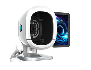
Portable 2025 Smart 3D Skin Analyzer Machine with AI Facial Diagnostics
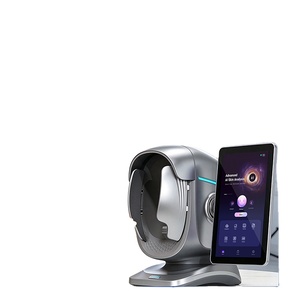
AI Skin Analyzer 7-Inch Touchscreen Scanner (36MP Optics & Multi-Language)
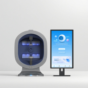
X5 Pro 8-Spectrum Clinical Skin Analyzer for Pigmentation & Moisture Detection

2025 Facial Scanning Skin Analysis Beauty Device for Pigmentation & Acne
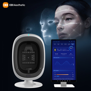
2025 Digital 3D Face Pigmentation Tester & Spectrum Diagnostic Camera
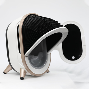
3D Smart Magic Mirror Skin Analyzer System for Salon Equipment Supplies
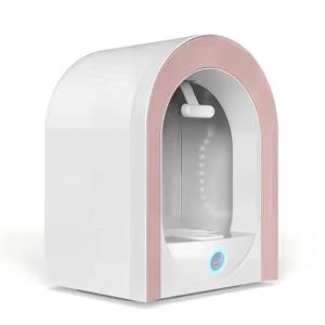
Professional AI Facial Skin Analyzer for Melasma & Deep Pigmentation
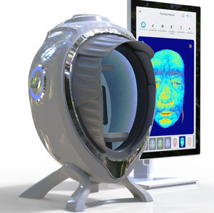
Wholesale Full Face Spectrum Skin Analyzer Machine for Deep Freckle Detection
Multi-Spectral Physics in Modern Dermatological Diagnostics
Dermatological consultations in contemporary aesthetic medicine have evolved beyond subjective visual evaluations. As an established Spectrum Skin Diagnostic Camera Manufacturer & Supplier serving Sri Lanka, our engineering core centers on the optical physics of light-tissue interaction. Human skin is a complex, multi-layered biological matrix comprising the epidermis, dermo-epidermal junction (DEJ), papillary dermis, and reticular dermis. Light rays at differing wavelengths exhibit variable absorption, scattering, and penetration depth profiles across these anatomical strata.
Standard white light (RGB daylight) reflects primarily off the stratum corneum, masking underlying cellular structures due to surface glare and diffuse reflection. To bypass this barrier, our multi-spectral imaging devices deploy proprietary optical filters and synchronized light-emitting arrays across distinct light spectra:
Cross-Polarized Light
Eliminates superficial specular reflections, allowing light to penetrate into the papillary dermis. Reveals subsurface vascular networks, telangiectasia, erythema, and underlying inflammation.
Parallel-Polarized Light
Enhances surface topography, fine lines, structural wrinkles, pore morphology, and micro-relief irregularities across the epidermal layer.
365nm UV Fluorescence
Excites fluorophores within cutaneous tissue. Detects active Cutibacterium acnes porphyrins, subterranean sun damage, and compromised lipid barrier function.
True Wood's Light Spectrum
Essential for differentiating epidermal melasma (which fluoresces brightly) from dermal melasma (which shows reduced contrast), guiding precise chemical peel selection.
Brown Spectrum Algorithm
Isolates hyper-pigmentation density mapping, quantifying deep melanin clusters before they manifest visibly on the cutaneous surface.
Red Spectrum Mapping
Extracts hemoglobin concentrations to track rosacea, acne flare-up zones, post-procedure recovery, and vascular neogenesis.
Information Gain Principle: By capturing 10 distinct spectral datasets in less than 3 seconds, our diagnostic systems generate quantitative optical density scores, removing clinical guesswork and creating an undeniable visual record for patient consultations.
Equatorial Tropical Dermatology: Sri Lanka Market Analysis & Phototype Dynamics
Operating as a premium Spectrum Skin Diagnostic Camera Supplier serving Sri Lanka requires deep localization rooted in regional dermatological reality. Sri Lanka is situated in an equatorial maritime zone (Latitude 5°55' to 9°51' N), exposing its population to intense solar irradiance with peak UV Index (UVI) values frequently exceeding 11–12 year-round in cities like Colombo, Kandy, Galle, and Jaffna.
The predominant cutaneous demographics in Sri Lanka encompass Fitzpatrick Phototypes IV, V, and VI. These melanin-rich phototypes present unique clinical challenges. While darker skin provides inherent photoprotection against UV-induced carcinogenesis, it possesses hyper-reactive melanocytes that predispose patients to higher incidences of Post-Inflammatory Hyperpigmentation (PIH), stubborn Melasma, Solar Lentigines, and lichen planus pigmentosus.
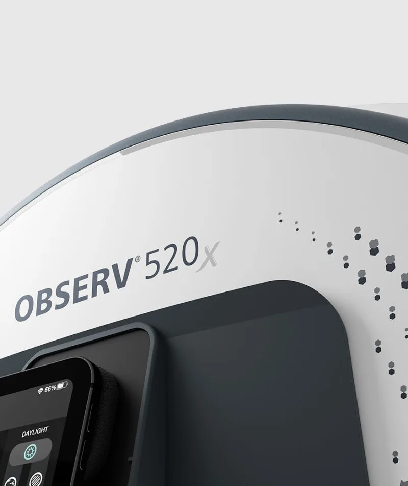
Furthermore, Sri Lanka's high relative humidity (75%–90%) triggers elevated sebum excretion, follicular occlusion, and widespread Malassezia folliculitis. When dermatologists or aesthetic physicians in Colombo attempt invasive procedures—such as Q-switched Nd:YAG laser toning, chemical peeling, or micro-needling RF—without baseline spectral mapping, the risk of triggering severe secondary PIH is exceptionally high.
| Diagnostic Parameter | Standard RGB Inspection | Our Multi-Spectrum AI Camera System | Clinical Advantage for Sri Lankan Clinics |
|---|---|---|---|
| Melasma Depth Classification | Surface Only | Epidermal / Dermal / Mixed Analysis | Prevents inappropriate laser fluence settings on deep dermal melasma. |
| Sub-Clinical UV Damage | Invisible | 365nm UV Fluorescence Isolation | Visualizes hidden melanin clusters before visible manifestation. |
| Sebum & Porphyrin Density | Qualitative Estimate | Quantitative Porphyrin Pixel Count | Differentiates bacterial acne from humidity-induced folliculitis. |
| Vascular Erythema (PIH Risk) | Masked by Melanin | Cross-Polarized Red Hemoglobin Separation | Identifies underlying inflammation in Fitzpatrick Types IV-VI. |
Localized Application Scenarios across Sri Lankan Aesthetic Centers
Our spectrum diagnostic cameras are engineered to address the specific workflow and commercial requirements of modern Sri Lankan practitioners:
-
Scenario A: Pre-Laser Safety Protocols in Colombo 03 & 07 Clinics
Aesthetic practitioners in premier Colombo clinics utilize our cross-polarized and UV spectrum modes to screen patients prior to energy-based device (EBD) therapies. By quantifying baseline inflammation and epidermal vs. dermal melanin ratio, doctors can set safer laser parameters, avoiding post-treatment hyperpigmentation lawsuits and enhancing patient trust.
-
Scenario B: Medi-Spa Hydration & Barrier Monitoring in Coastal Galle & Negombo
Luxury wellness resorts and medi-spas catering to international tourists and affluent locals in coastal regions use parallel-polarized imaging to evaluate lipid barrier breakdown caused by saltwater exposure and intense tropical humidity, recommending custom restorative regimens backed by digital proof.
-
Scenario C: Counterbalance to Unregulated Whitening Creams
Due to the prevalence of over-the-counter hydroquinone and mercury-tainted bleaching formulations in South Asia, dermatologists encounter severe cutaneous toxicity, exogenous ochronosis, and thinned skin barriers. Our spectrum cameras visually document epidermal atrophy and vascular damage under UV and red spectrums, convincing patients to halt toxic regimens and adopt medical treatments.
-
Scenario D: Longitudinal Treatment Verification in Kandy Clinical Centers
Dermatology clinics in Kandy employ our automatic 3D head positioners and standard illumination geometry to capture pixel-perfect "Before & After" diagnostic scans across 3-month, 6-month, and 12-month care cycles, providing scientific proof of pigment clearance and anti-aging efficacy.
Frequently Asked Questions (FAQ) for Sri Lanka Procurement
Upgrade Your Sri Lankan Practice with Clinical Multi-Spectral Diagnostics
Join leading dermatologists and aesthetic clinics in Colombo, Kandy, and Galle who rely on our spectrum diagnostic cameras to enhance patient retention, prevent laser risks, and drive skincare sales.