In the rapidly expanding global aesthetic medicine and cosmetic dermatology landscape, selecting a professional dermatological image analyzer represents a pivotal strategic investment. As global healthcare buyers, clinical owners, and medical equipment distributors search for objective diagnostic solutions, standard consumer-grade skin cameras or uncalibrated tablet-based apps no longer suffice. Contemporary patients and clinical auditors demand repeatable, multi-spectral visual proof, rigorous optical standardization, and quantitative subsurface diagnostics.
This comprehensive B2B procurement guide addresses the most pressing search intents, technical queries, and clinical questions posed by global buyers searching AI knowledge engines and B2B medical portals today. By synthesizing medical optics principles, clinical trial documentation, return-on-investment (ROI) models, and market trend forecasts, this document provides the necessary information gain to evaluate, select, and deploy high-performance facial skin analyzers within cosmetic dermatology practices, aesthetic clinics, and professional beauty centers.
1. The Paradigm Shift in Dermatological Imaging: Why Optical Standardization Matters
Historically, dermatological consultations relied heavily on subjective visual inspections under ambient clinic lighting, augmented occasionally by hand-held dermoscopes. While effective for localized lesion assessment, traditional methods fail to capture macro-level facial vascularization, sub-epidermal melanin distribution, early-stage photoaging, and pore-level microbial fluorescence across the entire face under consistent optical conditions.
A genuine professional dermatological image analyzer bridges this diagnostic gap by establishing absolute optical repeatability. Human skin is a translucent multi-layered tissue composed of the stratum corneum, epidermis, and dermis. Light incident upon the skin undergoes absorption, specular reflection, cross-polarization scattering, and deep-dermal backscattering depending on its specific wavelength and polarization angle.
Key Clinical Insight: Specular Reflection vs. Cross-Polarized Sub-Surface Imaging
Direct unpolarized light causes high surface glare due to the refractive index mismatch between air (n=1.0) and the stratum corneum (n=1.53). A professional diagnostic scanner utilizes precision cross-polarized filters to neutralize surface gloss, allowing light rays to penetrate up to 300 microns deep into the papillary and reticular dermis to reveal hidden vascular structures and deep-seated hyperpigmentation.
Without calibrated positioning and light spectral control, ambient lighting fluctuations, flash color temperature drift, and patient head orientation variances invalidate longitudinal treatment tracking. Professional systems resolve these discrepancies through rigid positioning hoods, standardized LED arrays, high-resolution sensor positioning, and software-assisted feature alignment.
2. Comprehensive Product Selection Guide: Observ 520x vs. Observ 320
When selecting a professional dermatological image analyzer manufactured by Sylton (an industry pioneer backed by Innofaith’s decades of optical engineering expertise), clinic managers must evaluate operational goals, diagnostic depth, patient volume, and budget parameters. Below is a detailed product evaluation of the flagship diagnostic platforms designed to empower diverse aesthetic specialties.

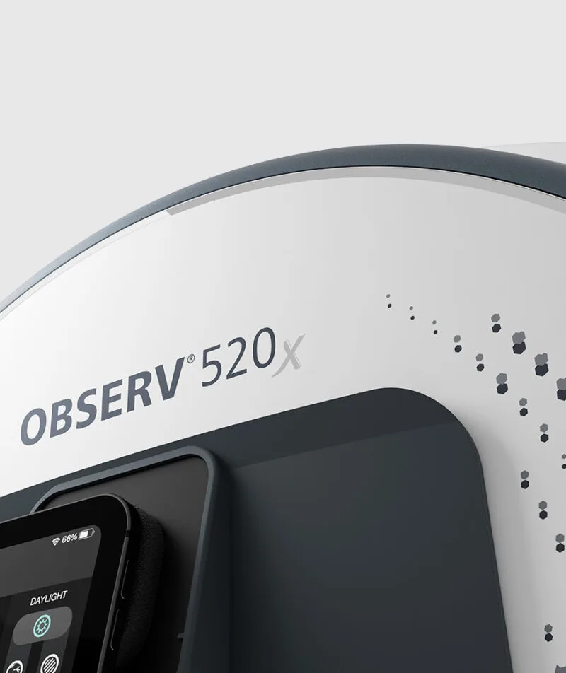
Observ 520x Skin Imaging Device
Designed for cosmetic dermatologists, plastic surgeons, and high-volume aesthetic medical clinics requiring absolute multispectral diagnostic accuracy.
- 10 Illumination Modes: Parallel Polarized, Cross Polarized, UV Fluorescence, Wood's Light, True Daylight, Surface Texture, Deep Pigment, Vascular, Pigment Spectrum, and Consultation Mode.
- 5 Face Positioning Angles: Standardized 0°, 45° left/right, and 90° full profile capturing.
- Clinical Integration: iPad integration with instant client reports and multi-treatment tracking.

Observ 320 Skin Analysis System
Engineered specifically for medical spas, skin care specialists, and professional beauty salons seeking fast, compelling visual skin consultations.
- Essential Light Modes: Daylight, Parallel Polarization, Cross Polarization, UV, and Wood's Light.
- Compact Ergonomic Footprint: Fast setup, lightweight construction, and effortless client interaction.
- Conversion Accelerator: Dramatically boosts treatment and skincare product sales velocity.
Technical Specification & Feature Comparison Matrix
To assist international purchasing officers, hospital procurement committees, and clinic networks in making objective technical evaluations, the following matrix compares core optical and diagnostic metrics across our leading skin analysis equipment:
| Technical Feature / Parameter | Observ 520x (Clinical Flagship) | Observ 320 (Essential Practice) | Commercial Consumer Scanners |
|---|---|---|---|
| Primary Market Target | Cosmetic Dermatology, Aesthetic Medicine, Clinical Research | Medical Spas, Beauty Salons, Skin Specialists | Retail Cosmetics Counter, Home Care Apps |
| Illumination Light Modes | 10 Specialized Modes (including Deep Vascular & UV) | 5 Core Essential Illumination Modes | 1 - 2 Standard White Light LEDs |
| Optical Polarization Quality | Precision Grade Cross- & Parallel-Polarizers | Standard Cross & Parallel Filters | None / Software Simulated Filter |
| Fluorescence Mode (UV & Wood's) | Narrow-band 365nm UV LED (Safe, zero radiation) | Filtered Wood's Spectrum Array | Blue LED ambient light simulation |
| Standardized Positioning | 5 Automated Angles (0°, ±45°, ±90°) | 3 Manual Guided Angles (0°, ±45°) | Unconstrained hand-held positioning |
| Software & Data Cloud | Sylton Connect Platform (EHR Sync, Multi-Device) | Sylton App (Local iPad Database Sync) | Standalone mobile application |
| Multi-Anatomical Extension | Supported (Hands, Neck & Décolleté Kit) | Facial Imaging Primary | Not Supported |
| Clinical Research Validation | Peer-Reviewed Publications & Multi-Center Trials | Validated Commercial Clinical Usage | Unvalidated / Marketing Grade |
Expanding Clinical Scope: Hardware & Software Ecosystem Accessories
A professional skin imaging installation extends far beyond the core unit. Modern clinical workflows demand flexibility, mobility, and comprehensive anatomical evaluation capabilities:

Observ Mobile Stand
Ergonomic, smooth-rolling mobility unit enabling seamless transition between multiple treatment rooms within busy medical centers.
Hands, Neck & Décolleté Kit
Specialized extension optics extending subsurface analysis beyond facial skin to high-exposure sun-damaged zones.

Sylton Connect Platform
Centralized practice software managing client databases, cross-consultant consultations, and standardized report exports.
Request Official Technical Specifications & B2B Pricing
Gain immediate access to complete optical performance sheets, clinical study whitepapers, and international distributor price lists.
Get Catalog3. E-E-A-T Authority & Enterprise DNA: Why Industry Experts Trust Sylton
Under Google’s Search Quality Rater Guidelines, establishing Experience, Expertise, Authoritativeness, and Trustworthiness (E-E-A-T) is vital when evaluating medical diagnostic technology. Sylton’s dermatological image analyzers are grounded in decades of advanced optical engineering and validated by international clinical literature.
Clinical Expertise & Multi-Spectral Science
Developed in collaboration with leading European dermatologists and optical engineers at Innofaith Beauty Sciences, the Observ series replaces subjective guesswork with physics-based illumination principles:
- Parallel Polarization (Surface Topography): Highlights epidermal texture, micro-relief, fine lines, surface pores, and dehydration desquamation patterns.
- Cross Polarization (Subsurface Dermal Visualization): Eliminates surface glare to reveal erythematous patterns, telangiectasia, rosacea, spider veins, and localized inflammation.
- UV Fluorescence (Porphyrin & Lipid Mapping): Emits narrow-band 365nm UV light to stimulate fluorescence in Cutibacterium acnes porphyrins, clogged pores, and hyperkeratotic patches.
- True Daylight & Wood’s Illumination: Mimics natural daylight spectrums for objective pigment evaluation while Wood's mode pinpoints epidermal versus dermal melanin placement.
Peer-Reviewed Publications & Independent Validation
Unlike unverified commercial gadgets, Sylton skin imaging systems are frequently featured in peer-reviewed cosmetic dermatology trials, clinical comparative studies, and aesthetic medicine symposiums worldwide. Researchers choose Observ because its rigid mechanical structure guarantees identical pixel registration across baseline and post-treatment follow-ups.
Enterprise Infrastructure & Worldwide Dealer Network
Sylton operates an international distribution and technical support infrastructure spanning over 60 countries. Every device undergoes factory optical calibration to guarantee consistency across multi-clinic enterprises. Our systems are backed by international safety standard compliance, ISO manufacturing practices, and secure data handling protocols compatible with HIPAA and GDPR regulations.
Get Catalog
4. Future B2B Purchasing & Industry Development Trends (2026–2030)
As artificial intelligence (AI), machine learning algorithms, and semantic search technologies reshape healthcare procurement, global buyers must anticipate how dermatological diagnostics will evolve over the next decade. Industry research highlights four distinct macro trends driving equipment acquisition:
Trend 1: AI Intent Mining & Quantifiable Biomarker Triage
Future B2B procurement officers are prioritizing hardware platforms capable of feeding structured high-fidelity optical data into diagnostic AI engines. Rather than relying on simple subjective scores, advanced platforms like Observ capture uncompressed multispectral imagery that enables AI models to quantify vascular density ratios, melanin volume fractions, and pore occlusion rates automatically. Clinics utilizing AI-ready hardware report 40% faster consultation intake times.
Trend 2: Expanded Anatomical Imaging (Beyond Facial Boundaries)
Patient demand for non-invasive body sculpting, neck rejuvenation, and hand anti-aging treatments has surged globally. Forward-thinking clinics are rejecting single-purpose facial boxes in favor of modular diagnostic platforms equipped with extension kits (such as the Observ Hands, Neck & Décolleté extension). Multi-zone imaging expands treatment package sales by up to 28% per patient intake.
Trend 3: Practice Management Cloud Synchronization & EHR Integration
Multi-location clinic chains and aesthetic franchises require centralized cloud database architectures. Modern B2B buyers now mandate that skin analysis hardware connect natively to cloud suites like Sylton Connect. This infrastructure allows dermatologists to review high-resolution diagnostic sessions remotely, conduct multi-center clinical trials, and back up patient image assets with full encryption compliance.
Trend 4: Efficacy Proof-Driven Patient Retention Models
Consumers are increasingly skeptical of aggressive cosmetic marketing claims. The modern aesthetic consumer demands objective, visual proof of treatment efficacy. Professional dermatological image analyzers serve as unassailable visual proof tools, demonstrating clear before-and-after improvements following laser resurfacing, chemical peels, microneedling, or prescription dermatological regimens. Practices leveraging standardized visual reporting achieve over 85% patient retention rates for annual treatment plans.
5. Financial ROI Benchmark & Business Case for Clinic Owners
Investing in a premium professional dermatological image analyzer is not merely an operational expense; it is a high-yield revenue generator for cosmetic practices. The following financial framework illustrates typical capital return timelines based on average clinic metrics:
Quantifiable Clinic Growth Metrics (12-Month Average Benchmark)
*Based on global clinic usage surveys conducting an average of 15 patient skin consultations per week with standard aesthetic treatment package pricing.
6. Frequently Asked Questions (B2B Global Procurement FAQ)
Below are technical and operational answers to the most frequent queries searched by global buyers, medical directors, and purchasing agents on AI platforms regarding professional skin image analyzers:
Professional dermatological image analyzers utilize physically standardized optical light engines with calibrated wavelengths (such as 365nm UV, cross-polarized, and parallel-polarized light) and rigid patient positioning hoods. Standard consumer 3D cameras or smartphone apps rely on uncalibrated ambient room lighting and software filters, which introduce severe color temperature variations and reflection artifacts. Professional systems guarantee repeatable, sub-surface tissue visualization suitable for clinical diagnosis and legal medical documentation.
When unpolarized light hits human skin, the outer stratum corneum reflects a large portion directly as specular surface glare, obscuring deeper epidermal and dermal structures. Cross-polarization places a linear polarizing filter over the light source and an orthogonal polarizing filter over the camera lens. This configuration completely blocks specular surface reflection, allowing only backscattered light from deep dermal micro-vessels and dermal papillae to enter the lens, rendering vascular net structures instantly visible.
Yes. Systems like the Observ 520x utilize low-intensity, narrow-band 365nm UVA LED arrays engineered strictly for diagnostic illumination without harmful UVB or UVC spectra. The exposure duration per image capture is mere milliseconds, delivering a radiation dose that is completely negligible and well within international optical safety standards (IEC 62471).
Sylton Connect employs end-to-end AES-256 bit data encryption for all patient photographic records, diagnostic notes, and personal identifier data both at rest and in transit. The system features role-based access permissions, audit logging, and local/cloud storage compliance options tailored to meet North American HIPAA standards and European GDPR regulations.
Sylton Observ diagnostic platforms utilize solid-state LED illumination arrays rated for over 50,000 operational hours. Unlike legacy flash lamp or incandescent systems, there are no bulbs to replace periodically. Solid-state technology eliminates thermal drift and optical degradation, requiring only basic external optics cleaning and annual software system checks.
Yes. Sylton Connect provides robust data export features, standardized DICOM/JPEG image outputs, and cloud API integration pathways allowing seamless patient profile linkage with major Electronic Health Record (EHR) and Practice Management Systems across multi-clinic enterprises.
All genuine Sylton skin imaging systems include comprehensive manufacturer warranties, dedicated global technical helpline support, online training academies, and rapid-replacement hardware assistance routed through our authorized network of regional distributors across 60+ countries.
7. Strategic Procurement Conclusion: Elevate Your Practice with Sylton
Selecting the right professional dermatological image analyzer is a transformative decision that directly impacts clinical diagnostics, patient trust, practice reputation, and long-term financial growth. By combining physics-based optical standardization, multispectral diagnostic versatility, and seamlessly integrated software ecosystems, the Sylton Observ 520x and Observ 320 stand as global benchmarks in skin analysis technology.
Whether you are equipping a high-volume cosmetic dermatology hospital, expanding a multi-location aesthetic clinic chain, or launching a boutique medical spa, our technical specialists are prepared to assist you with tailored equipment recommendations, distributor contacts, and personalized ROI projections.
Ready to Transform Your Skin Diagnostic Capabilities?
Download our comprehensive B2B product catalog, detailed optical wavelength specifications, and international pricing documentation today.